31 Jul, 2026
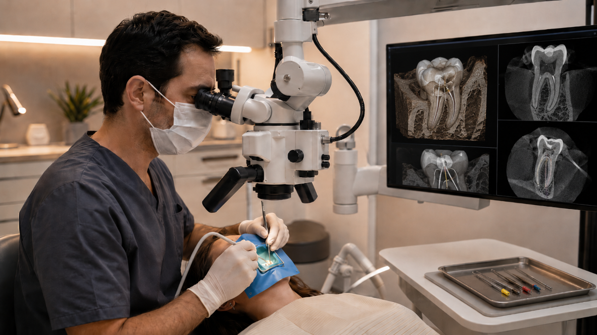
In surgical micro-endodontics, the operating microscope has become an everyday working tool. Micro-endodontics is the branch of endodontics that uses optical magnification, through the operating microscope, to work on root canals: structures that are often narrow, curved or calcified, and difficult to see with the naked eye or with traditional optical aids.
This is the premise behind the postgraduate program in Innovative Micro-Endodontics: Advanced Digital and Microscopic Techniques at Vita-Salute San Raffaele University (UniSR), designed for dentists who want to bring the operating microscope and digital technologies into their own clinical practice. Directed by Prof. Giuseppe Cantatore, the program runs for 12 months and carries 60 ECTS credits.
The curriculum integrates Cone Beam Computed Tomography (CBCT), which produces three-dimensional images of dental anatomy, a range of artificial intelligence software, and guided endodontics. Clinical training on real patients complements these technologies.
When the Microscope Becomes the Working Standard
In everyday dental practice, the operating microscope is almost always reserved for the most complex cases. The program reverses that habit. Each participant is trained to carry out an entire endodontic treatment, from diagnosis to canal filling, under optical magnification. The microscope appears from the first week of the course and stays central through the final clinical placement, with a module dedicated to operator positioning in both orthograde and surgical endodontics.
Direct visibility of the root canals reduces the margin of error during cleaning and shaping, particularly in anatomically complex cases. For a practicing clinician, this means a real shift in approach: every treatment is planned and carried out differently, from orthograde retreatments to endodontic surgery, where precision in the operating field matters even more.
Why Use the Microscope in Micro-Endodontics
A meta-analysis published in the Journal of Endodontics in 2010 compared traditional endodontic surgery, performed without optical aids, with microscope-assisted endodontic surgery: the success rate rose from 59% to 94%, a statistically significant difference. A more recent 2025 study confirms the same pattern outside surgery too: across more than 600 teeth treated with root canal therapy, outcomes under the microscope were significantly better than those achieved without optical magnification. It comes as no surprise that microscope use among endodontists rose from 52% to 90% in the United States between 1999 and 2008, and today exceeds 91% globally.
Inside the Micro-Endodontics Curriculum
The curriculum covers the full clinical pathway of modern endodontics, from diagnosis to complication management. It starts with pulpal and periapical pathology and pulp regeneration and revascularization techniques, then moves to canal irrigation with sodium hypochlorite, EDTA, and newer-generation activation systems.
A substantial part of the program is dedicated to nickel-titanium instrumentation systems, from rotary to reciprocating systems. The two kinematics behave differently in curved or calcified canals, and knowing both is essential.
The course also addresses what happens when the first treatment isn't enough: retreatments, removal of old posts and metal carriers, and management of fractured instruments and vertical fractures. One module is dedicated to endodontic microsurgery and laser use. The curriculum closes with bioceramic materials, used mainly in the most delicate cases, such as teeth with immature apices or roots undergoing resorption.
Digital Technologies and Artificial Intelligence in Endodontics
Endodontic planning is being transformed by new technologies and artificial intelligence. Cone beam computed tomography produces three-dimensional images of dental anatomy and can be paired with AI software for diagnostic analysis. One module in the program teaches how to minimize imaging artifacts and reduce the patient's radiation exposure.
Part of the course is dedicated to guided endodontics, a technique that combines DICOM files from CBCT scans with STL files produced by intraoral scanners. Overlaying the two datasets generates 3D-printed guides, used to precisely direct treatment in both orthograde and surgical endodontics. The program also takes a hands-on approach to this, with a module dedicated to the "print & try" technique, from scan to printed tooth.
Where Clinical Practice Happens
Hands-on training in the Micro-Endodontics program takes place on two levels. In the Smiley Lab, UniSR's dental simulation facility, each participant works at an individual station, practicing on advanced anatomical models before treating patients. This is where manual skill under the microscope is built, in a protected setting ahead of clinical practice.
The next step takes place in the Smart Clinics of the San Donato Group, where clinical training takes place with real patients under direct supervision. The expected commitment is around ten days, scheduled in agreement with the program's directors. The simulator-to-patient sequence transfers bench skills to an authentic clinical setting, one step at a time.
Program Structure: Duration, Credits, and Commitment
The program runs for 12 months and carries 60 ECTS credits, with admission reserved for candidates who pass a credentials review and an interview. Classes are taught in Italian, organized into 7 course weeks spread across the year: 5 in person, with lectures, workshops and live demonstrations, and 2 online, dedicated to clinical case discussions, research, and live seminars. A minimum attendance of 75% is required for both in-person teaching and other training formats.
Beyond the course weeks, the program includes clinical training and a final dissertation, which together account for nearly half of the 60 total ECTS credits: the largest single component of the program. Start dates and application deadlines are updated each year on the official course page.
How to Apply to the Micro-Endodontics Program
Admission requires a credentials review and an interview. Requirements, costs, and updated application deadlines are published on the official course page on the UniSR website, along with the detailed curriculum and activity calendar.
Bibliography
Setzer F.C., Shah S.B., Kohli M.R., Karabucak B., Kim S., "Outcome of endodontic surgery: a meta-analysis of the literature, part 1: comparison of traditional root-end surgery and endodontic microsurgery", Journal of Endodontics, 2010;36(11):1757-1765. https://pubmed.ncbi.nlm.nih.gov/20951283/
"Effectiveness of microscope-assisted root canal treatment in permanent posterior teeth: A retrospective cohort study", International Endodontic Journal, April 2025. https://www.sciencedirect.com/science/article/abs/pii/S0300571225002155
American Association of Endodontists, "The Dental Operating Microscope", Colleagues for Excellence, Winter 2016. https://www.aae.org/specialty/wp-content/uploads/sites/2/2017/07/winter2016microscopes.pdf
Cheung M.C. et al., "Global survey of endodontic practice and adoption of newer technologies", International Endodontic Journal, 2023. https://onlinelibrary.wiley.com/doi/abs/10.1111/iej.13982
Written by

UniSR Communication Team
Thanks to the contribution of the various team members, the UniSR Marketing and Communications Service deals with the multiple communication areas of the University: news scouting, creation of news, audio and video, event organization, website management and institutional social media, drafting and publication of newsletters, support for institutional relations. The Service interacts with all the main stakeholders (students, teachers, technical and administrative staff, research community, territory) in order to support and potential communication (internal and external) of the initiatives related to teaching, research and public engagement.
Visit the author's page