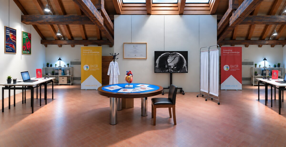
From 1 to 11 October, students and visitors can explore AI in medical diagnosis by stepping into a doctor’s shoes. They can do so at Spazio Viterbi in Bergamo with HumAIn Medicine, the app developed by Vita-Salute San Raffaele University in Milan for BergamoScienza 2026. During a one-hour lab session, participants take charge of three cardiovascular clinical cases, analyse the clinical data and diagnostic tests, and assess the algorithm’s results to reach a clinical decision.
The app is based on CORO-CTAIOMICS, a research project run by UniSR and IRCCS San Raffaele Hospital and funded by Italy’s National Recovery and Resilience Plan (PNRR). The project uses artificial intelligence to analyse coronary CT angiography (CCTA) images and estimate the risk of major cardiovascular events in the four years following the scan. For the lab, the research tools were adapted into an educational app designed for the general public.
How HumAIn Medicine Works
The app simulates a patient’s assessment in a cardiology clinic and guides the public through several stages of clinical practice, starting with the patient’s history. The first figure participants interpret is a score recommended by the European Society of Cardiology guidelines and used to estimate the likelihood that the patient has significant narrowing of the coronary arteries. The calculation takes into account age, sex, symptoms (such as the nature of the chest pain and any shortness of breath) and cardiovascular risk factors including high blood pressure, diabetes, smoking and abnormal blood lipid levels. This value helps the cardiologist decide whether further tests are needed, such as a diagnostic radiology scan called coronary CT angiography. Visitors then open the CCTA scan, a non-invasive test that uses X-rays and a contrast agent to produce detailed images of the arteries that supply blood to the heart. The images show any plaques or narrowing in the coronary arteries.
At this point, participants grade the scan using CAD-RADS, the classification system that standardises how the presence and severity of coronary artery disease are reported on CT. Scores range from 0 (no plaque or stenosis) to 5 (total occlusion of at least one coronary artery).
Finally, those trying the app review the results of the algorithm, which analyses the same CCTA scan and returns quantitative information on tissue characteristics. This information complements the clinician’s own reading of the scan.
For those who want to find out more, the lab, set up as a cardiologist’s consulting room, hosts a series of panels that show and explain the physics behind the main imaging techniques, from X-rays and ultrasound to CT, PET and MRI.
AI in Medical Diagnosis: What the Algorithm Sees
When reviewing a CCTA scan, the radiologist evaluates the coronary arteries and looks for abnormalities, such as plaques or narrowing that can restrict blood flow to the heart. Yet the images contain far more data than even the most experienced human eye can interpret. CORO-CTAIOMICS, the research project behind the app, works on precisely this additional data.
How CORO-CTAIOMICS Works
At the hospital, the scan is uploaded to the research platform, which analyses it automatically. The algorithms first identify the heart and the coronary arteries. They then examine the area around them, which includes the arterial wall, any plaques and the surrounding adipose tissue. This technique, known as radiomics, extracts thousands of quantitative parameters from the images that describe tissue characteristics such as shape, intensity and organisation.
The fat surrounding the coronary arteries, known as pericoronary fat, is a metabolically active tissue that can reflect changes in the environment around the arteries. For this reason, the research project treats the coronary wall, any plaques and the surrounding fat as a single region and measures its characteristics quantitatively, gathering information that helps build a clearer picture of cardiovascular risk.
From Diagnosis to Cardiovascular Risk
CORO-CTAIOMICS pairs the image assessment with a prediction of future cardiovascular risk. The algorithms combine the figures derived from the images with some of the patient’s clinical data, such as age, sex and main risk factors. Within a few minutes, the platform returns an individual estimate of cardiovascular risk, together with colour maps showing how certain quantitative parameters extracted by the algorithm vary across the regions analysed. In the HumAIn Medicine app, participants see these results in a version adapted for the public and base their decisions on them.
The prediction is clinically valuable, yet it remains a probability rather than a certainty. According to the Istituto Superiore di Sanità, the Italian National Institute of Health, cardiovascular diseases are the leading cause of death in Italy and can develop for years without obvious symptoms. Identifying the patients most at risk early on is one of the major challenges in medicine. The research project aims to draw more information from a routine CCTA scan to support clinical assessment. IRCCS San Raffaele Hospital’s S-RACE platform, designed to bring predictive models from research to the bedside, shares this goal.
Colour Maps: The Doctor Makes the Call
In the HumAIn Medicine educational app, the AI results also appear as colour maps overlaid on the image of the proximal segments of the coronary arteries. A red area instinctively suggests danger. The legend makes clear that the colour indicates the value of that specific parameter and where it falls on the relevant scale, rather than the level of risk itself. To understand whether that variation affects risk, the map has to be read together with the anatomical image, the clinical data and the algorithm’s other results.
This synthesis is the doctor’s job, and the lab created by UniSR lets the public try it first-hand. In clinical practice, the algorithm produces figures and maps in a matter of minutes. Deciding what they mean for that particular person, and which treatment to offer, calls for clinical judgement.
In this way, the lab gives a concrete picture of the role AI can play in clinical practice: a tool that supports professionals by offering new information without replacing their judgement and responsibility.
Some ethical questions remain open. An algorithm trained on biased or incomplete data may produce less accurate results for groups of patients under-represented in the data used to develop it, with possible consequences for clinical assessment. We explored this issue in our article on bias in medical AI. Health data is particularly sensitive, and its use is subject to specific rules on data protection, privacy and ethics.
The Team Behind HumAIn Medicine
HumAIn Medicine is the work of UniSR’s Public Engagement and Research Communication Office, which turned selected content from a real research project into an educational experience.
It was developed in collaboration with the Advanced Imaging for Personalized Medicine Unit and Prof. Antonio Esposito, Full Professor of Radiology and President of the Master’s Degree in Health Informatics, building on the content and research tools of the CORO-CTAIOMICS project.
During the festival, the lab is run by BergamoScienza volunteers and by students from MUSA, UniSR’s student group dedicated to science communication.
Visitors can try HumAIn Medicine until 11 October. Participation is free, but booking via the BergamoScienza website is required. Teachers can also book for their classes there.
Want to explore the relationship between AI and medicine further? Read our article on AI in scientific research.